- März 1, 2021
IMAGING AND DIAGNOSIS FOR PLANNING THE SURGICAL PROCEDURE
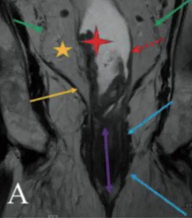
The preoperative imaging diagnosis of rectal cancer lies at the heart of oncological staging and has a crucial influence on patient management and therapy planning. Rectal cancer is common, and accurate preoperative staging of tumors using high-resolution magnetic resonance imaging (MRI) is a crucial part of modern multidisciplinary team management (MDT). Indeed, rectal MRI has the ability to accurately evaluate a number of important findings that maBay impact patient management, including distance of the tumor to the mesorectal fascia, presence of lymph nodes, presence of extramural vascular invasion (EMVI), and involvement of the anterior peritoneal reflection/peritoneum and the sphincter complex. Many of these findings are difficult to assess in non-expert hands. In this chapter, we present currently used staging modalities with focus on MRI, including optimization of imaging techniques, tumor staging, interpretation help as well as essentials for reporting.
This medical abstract is published in the following media:
IntechOpen © 2020
World Journal of Gastroenterology © 2004-2021
The full-length publication in English can be found at the following link:
English version: „Imaging and Diagnosis for Planning the Surgical Procedure“
The full-length publication in Hungarian can be found at the following link:
Hungarian version: „Imaging and Diagnosis for Planning the Surgical Procedure“